

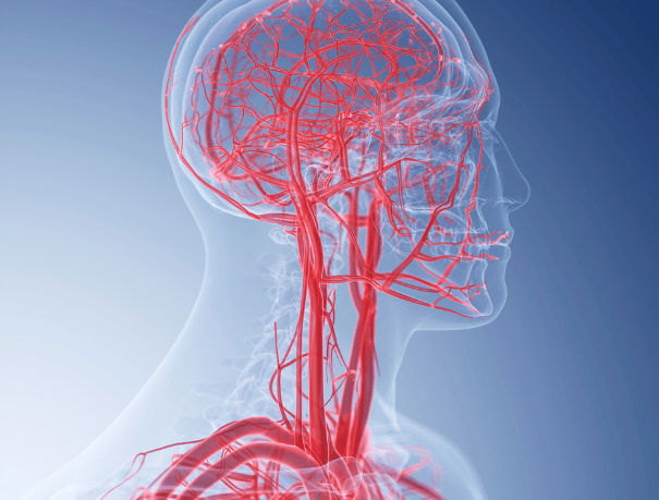
MRI Angiography (ARM)
What does it consist of?
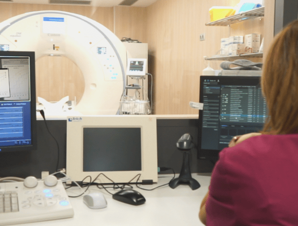
Unlike traditional angiography, which involves placing a tube (catheter), MRI Angio (MRA) is performed by administering intravenous contrast. To carry out the test, you will have to lie face up with your head resting on a cushion and a radiologist will inject you with contrast to mark the area to be studied. Next, the stretcher will move until the area of the body to be studied is in the center of the resonance equipment.
The test can last from 20 to 45 minutes. During this time, you will have to remain as calm as possible, since the taking of images is not continuous, but different image planes are made. Once finished you can continue with your daily life as normal.
CASES IN WHICH IT IS RECOMMENDED
Who is it for?
At Paracelso Sagasta we perform angioresonance or MRA of the different parts of the body:
- MRI angiography of the abdomen. Study of the vascular anatomy of the abdomen.
- MR angiography of the lower extremities. Explore the blood vessels in the legs.
- Hand MRI angiography. Evaluate the arteries of the hand in case of suspicion of vascular disease in the upper extremity.
- Angiography MRI standing. Study of the arterial supply of the foot.
- Renal MRI angiography. Evaluates the condition of the renal arteries, responsible for supplying blood to the kidney, adrenal gland, and ureters.
- Subclavian MRI angiography. Explore the subclavian artery, which, along with the carotid artery, is the main supplier of oxygen-rich blood to the head, brain, neck, and chest area.
- Angio MRI of carotids. Study of possible anomalies in the carotid arteries, located on both sides of the neck and the main supply routes of oxygenated blood to the brain.
- Angio MRI of the skull. Study of the vertebrobasilar system. This includes the vertebral arteries and the basilar artery, which supply blood to the posterior area of the brain and to both cerebral hemispheres.
- Chest MRI angiography. Study of the vascular anatomy of the thorax, with special emphasis on the thoracic aorta.
- Pelvic MRI angiography. Explore the iliac arteries, which supply oxygenated blood to the bladder, uterus and cervix, vagina and rectum, among other pelvic tissues such as the gluteal and perineal muscles.
INSTRUCTIONS
How should you prepare?
- Contrast administration: This type of exploration requires intravenous contrast administration.
- Metal objects: The technician will give you the necessary instructions, provide you with a gown and ask you to remove any metal objects (jewelry, watch, piercings, hairpins, mobiles, dental prostheses and hearing aids).
- Heart devices: It’s important to tell the technician about any devices you’re implanted in your body (pacemakers, electrodes, and clips from previous surgeries).
- Hydration and lactation: Drink plenty of water to eliminate contrast from the body. If you are breastfeeding, you should stop for 48 hours. You should discard breast milk.

THE SPECIALISTS WHO WILL ASSIST YOU IN PARACELSO SAGASTA
A team of professionals who take care of you


The opinion of experts
Paracelso Sagasta's blog
You will find advice from our professionals on how to improve your health and information on the latest technologies applied in the medical health sector.
 Health and advice
Make 2025 your healthiest year: a guide to positive habits
 Health and advice
10 Tips for a healthy Christmas
 Health and advice